ملف:Coronavirus. SARS-CoV-2.png

من أرابيكا، الموسوعة الحرة
اذهب إلى التنقل اذهب إلى البحث

الملف الأصلي(2٬048 × 2٬048 بكسل حجم الملف: 4٫54 ميجابايت، نوع MIME: image/png)

ملخص

الوصف
Deutsch: Wissenschaftlich genaues Atommodell der äußeren Struktur des SARS Coronavirus 2 (SARS-CoV-2), einem Stamm (genetische Variante) des Coronavirus, der die Coronavirus-Krankheit (COVID-19) verursachte und erstmals im Dezember 2019 in Wuhan, China, identifiziert wurde.

Jeder einzelne Ort (amorpher Fleck) ist ein Atom von:

 
kobalt: Virushülle
 
purpurrot: Hüllproteine
 
grün: Matrixproteine
 
orange: Glucose (Glycane)
 
türkis: Spike-Proteine
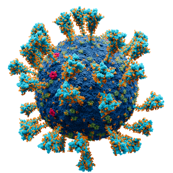
English: Scientifically accurate atomic model of the external structure of the Severe Acute Respiratory Syndrome CoronaVirus 2 (SARS-CoV-2), a strain (genetic variant) of the coronavirus that caused coronavirus disease (COVID-19), first identified in Wuhan, China, during December 2019

Each separate locus (amorphous blob) is a molecule of:

 
cobalt: membrane
 
crimson: E protein
 
green: M protein
 
orange: glucose (glycan)
 
turquoise : S (spike) glycoprotein
Español: Modelo atómico de la estructura externa del SARS-CoV-2. Cada "bola" es un átomo.
Leyenda:
 
azul cobalto — membrana
 
turquesa — glicoproteína de pico (S)
 
carmesí — proteína E
 
verde — proteína M
 
naranja — glucosa
Русский: Научно достоверная атомарная модель внешней структуры коронавируса (SARS-CoV-2). Каждый "шарик" — атом. Опубликовано на N+1.

Научный консультант:
Никитин Н. А. (доктор биологических наук, специалист в области вирусологии)
Борисевич С.С. к.х.н специалист по молекулярному моделированию поверхностных вирусных белков, руководитель группы «Кванты и Динамика», (с.н.с. лаборатория химической физики УфИХ РАН)
Архипова В.И. (специалитет по направлению фундаментальная и прикладная химия. Лаборатория химии РНК, Институт химической биологии и фундаментальной медицины Сибирского отделения Российской академии наук).

  • В работе были использованы следующие структуры из открытых источников:

Protein Data Bank: 2mls, 6y3y, 5x29, 6yyt Charmm-gui: 6vsb_1_1_1_S309

  • Цветовое обозначение:
 
кобальт — мембрана.
 
бирюза — S-белок.
 
малиновый — E-белок.
 
зелёный — M-белок.
 
оранжевый — гликаны.
Проект был сделан в соответствии с научными источниками:
  1. Surya, W., Li, Y., Torres, J. Structural model of the SARS coronavirus E channel in LMPG micelles. // Biochim. Biophys. Acta - Biomembr. – 2018. – Vol. 1860. – N. 6. – P. 1309–1317.
  2. Koppisetti, R. K., Fulcher, Y. G., Jurkevich, A., Prior, S. H., Xu, J., Lenoir, M., Overduin, M., Van Doren, S. R. Ambidextrous binding of cell and membrane bilayers by soluble matrix metalloproteinase-12. // Nat. Commun. – 2014. – Vol. 5. – P. 1–14.
  3. Hillen, H. S., Kokic, G., Farnung, L., Dienemann, C., Tegunov, D., Cramer, P. Structure of replicating SARS-CoV-2 polymerase. // Nature. – 2020. – Vol. 584. – N. 7819. – P. 154–156.
  4. Harris, L. J., Larson, S. B., Hasel, K. W., McPherson, A. Refined structure of an intact IgG2a monoclonal antibody. // Biochemistry. – 1997. – Vol. 36. – N. 7. – P. 1581–1597.
  5. Noreng, S., Bharadwaj, A., Posert, R., Yoshioka, C., Baconguis, I. Structure of the human epithelial sodium channel by cryo-electron microscopy. // Elife. – 2018. – Vol. 7. – P. 1–23.
  6. Almond, A., DeAngelis, P. L., Blundell, C. D. Hyaluronan: The Local Solution Conformation Determined by NMR and Computer Modeling is Close to a Contracted Left-handed 4-Fold Helix. // J. Mol. Biol. – 2006. – Vol. 358. – N. 5. – P. 1256–1269.
  7. Hurdiss, D. L., Drulyte, I., Lang, Y., Shamorkina, T. M., Pronker, M. F., van Kuppeveld, F. J. M., Snijder, J., de Groot, R. J. Cryo-EM structure of coronavirus-HKU1 haemagglutinin esterase reveals architectural changes arising from prolonged circulation in humans. // Nat. Commun. – 2020. – Vol. 11. – N. 1. – P. 1–10.
  8. Yan, Renhong, Yuanyuan Zhang, Yaning Li, Lu Xia, Yingying Guo, Q. Z. Structural basis for the recognition of SARS-CoV-2 by full-length human ACE2. // Science (80-. ). – 2020. – Vol. 3. – N. 3. – P. 1–8.
  9. Javitt, G., Khmelnitsky, L., Albert, L., Bigman, L. S., Elad, N., Morgenstern, D., Ilani, T., Levy, Y., Diskin, R., Fass, D. Assembly Mechanism of Mucin and von Willebrand Factor Polymers. // Cell. – 2020. – Vol. 183. – N. 3. – P. 717-729.e16.
  10. Daniel Wrapp, Nianshuang Wang, Kizzmekia S. Corbett, Jory A. Goldsmith, Ching-Lin Hsieh, Olubukola Abiona, B. S. G., McLellan, and J. S. Cryo-EM structure of the 2019-nCoV spike in the prefusion conformation. // Science (80-. ). – 2020. – Vol. 21. – N. 1. – P. 1–9.
  11. Wang, M. Y., Zhao, R., Gao, L. J., Gao, X. F., Wang, D. P., Cao, J. M. SARS-CoV-2: Structure, Biology, and Structure-Based Therapeutics Development. // Front. Cell. Infect. Microbiol. – 2020. – Vol. 10. – N. November. – P. 1–17.
  12. Yao, H., Song, Y., Chen, Y., Wu, N., Xu, J., Sun, C., Zhang, J., Weng, T., Zhang, Z., Wu, Z., Cheng, L., Shi, D., Lu, X., Lei, J., Crispin, M., Shi, Y., Li, L., Li, S. Molecular Architecture of the SARS-CoV-2 Virus. // Cell. – 2020. – Vol. 183. – N. 3. – P. 730-738.e13.
  13. Oostra, M., de Haan, C. A. M., de Groot, R. J., Rottier, P. J. M. Glycosylation of the Severe Acute Respiratory Syndrome Coronavirus Triple-Spanning Membrane Proteins 3a and M. // J. Virol. – 2006. – Vol. 80. – N. 5. – P. 2326–2336.
  14. B.W. Neuman, M. J. B. Supramolecular Architecture of the Coronavirus Particle. // Adv. Virus Res. – 2020. – Vol. 96. – P. 1–27.
  15. Neuman, B. W., Kiss, G., Kunding, A. H., Bhella, D., Baksh, M. F., Connelly, S., Droese, B., Klaus, J. P., Makino, S., Sawicki, S. G., Siddell, S. G., Stamou, D. G., Wilson, I. A., Kuhn, P., Buchmeier, M. J. A structural analysis of M protein in coronavirus assembly and morphology. // J. Struct. Biol. – 2011. – Vol. 174. – N. 1. – P. 11–22.
  16. Yu, A., Pak, A. J., He, P., Monje-Galvan, V., Casalino, L., Gaieb, Z., Dommer, A. C., Amaro, R. E., Voth, G. A. A multiscale coarse-grained model of the SARS-CoV-2 virion. // Biophys. J. – 2021. – Vol. 120. – N. 6. – P. 1097–1104.
  17. Yao, H., Song, Y., Chen, Y., Wu, N., Xu, J., Sun, C., Zhang, J., Weng, T., Zhang, Z., Wu, Z., Cheng, L., Shi, D., Lu, X., Lei, J., Crispin, M., Shi, Y., Li, L., Li, S. Molecular architecture of the SARS-CoV-2 virus. // Cell. – 2020. – Vol. 183. – N. 3. – P. 730–738.
  18. Choi, Y. K., Cao, Y., Frank, M., Woo, H., Park, S. J., Yeom, M. S., Croll, T. I., Seok, C., Im, W. Structure, Dynamics, Receptor Binding, and Antibody Binding of the Fully Glycosylated Full-Length SARS-CoV-2 Spike Protein in a Viral Membrane. // J. Chem. Theory Comput. – 2021. – Vol. 17. – N. 4. – P. 2479–2487.

Первичные источники:


Производные моделей протеинов созданы на базе моделей со свободной лицензией (freely available for both non-commercial and commercial use).
Українська: Атомарна модель зовнішньої структури коронавірусу SARS-CoV-2. Кожен «кулька» — атом.
التاريخ
المصدر

عمل شخصي. Scientific consultants:

Nikitin N.A., Doctor of Biological Sciences, Department of Virology, Faculty of Biology, Lomonosov Moscow State University.
Borisevich S.S. Candidate of Chemical Sciences, Specialist in Molecular Modeling of Viral Surface Proteins, Senior Researcher, Head of the "Quantum & Dynamics»,Laboratory of Chemical Physics, Ufa Institute of Chemistry RAS
Arkhipova V.I., specialization in Fundamental and Applied chemistry, senior engineer, RNA Chemistry Laboratory, Institute of chemical biology and fundamental medicine SB RAS
المؤلف Alexey Solodovnikov (Idea, Producer, CG, Editor), Valeria Arkhipova (Scientific Сonsultant)
الترخيص
(إعادة استخدام هذا الملف)

Published by N+1 a popular science online publication of Russia (https://nplus1.ru/), protein models are derivative works of the

free license site (freely available for both non-commercial and commercial use)
إصدارات أخرى

Sources

Primary sources:

The following structures from open sources were used in the work, Protein Data Bank (https://www.rcsb.org):

Additional sources:

  1. Surya, W., Li, Y., Torres, J. Structural model of the SARS coronavirus E channel in LMPG micelles // Biochim. Biophys. Acta - Biomembr. – 2018. – Vol. 1860. – N. 6. – P. 1309–1317.
  2. Koppisetti, R. K., Fulcher, Y. G., Jurkevich, A., Prior, S. H., Xu, J., Lenoir, M., Overduin, M., Van Doren, S. R. Ambidextrous binding of cell and membrane bilayers by soluble matrix metalloproteinase-12 // Nat. Commun. – 2014. – Vol. 5. – P. 1–14.
  3. Hillen, H. S., Kokic, G., Farnung, L., Dienemann, C., Tegunov, D., Cramer, P. Structure of replicating SARS-CoV-2 polymerase // Nature. – 2020. – Vol. 584. – N. 7819. – P. 154–156.
  4. Harris, L. J., Larson, S. B., Hasel, K. W., McPherson, A. Refined structure of an intact IgG2a monoclonal antibody // Biochemistry. – 1997. – Vol. 36. – N. 7. – P. 1581–1597.
  5. Noreng, S., Bharadwaj, A., Posert, R., Yoshioka, C., Baconguis, I. Structure of the human epithelial sodium channel by cryo-electron microscopy // Elife. – 2018. – Vol. 7. – P. 1–23.
  6. Almond, A., DeAngelis, P. L., Blundell, C. D. Hyaluronan: The Local Solution Conformation Determined by NMR and Computer Modeling is Close to a Contracted Left-handed 4-Fold Helix // J. Mol. Biol. – 2006. – Vol. 358. – N. 5. – P. 1256–1269.
  7. Hurdiss, D. L., Drulyte, I., Lang, Y., Shamorkina, T. M., Pronker, M. F., van Kuppeveld, F. J. M., Snijder, J., de Groot, R. J. Cryo-EM structure of coronavirus-HKU1 haemagglutinin esterase reveals architectural changes arising from prolonged circulation in humans // Nat. Commun. – 2020. – Vol. 11. – N. 1. – P. 1–10.
  8. Yan, Renhong, Yuanyuan Zhang, Yaning Li, Lu Xia, Yingying Guo, Q. Z. Structural basis for the recognition of SARS-CoV-2 by full-length human ACE2 // Science (80-. ). – 2020. – Vol. 3. – N. 3. – P. 1–8.
  9. Javitt, G., Khmelnitsky, L., Albert, L., Bigman, L. S., Elad, N., Morgenstern, D., Ilani, T., Levy, Y., Diskin, R., Fass, D. Assembly Mechanism of Mucin and von Willebrand Factor Polymers // Cell. – 2020. – Vol. 183. – N. 3. – P. 717-729.e16.
  10. Daniel Wrapp, Nianshuang Wang, Kizzmekia S. Corbett, Jory A. Goldsmith, Ching-Lin Hsieh, Olubukola Abiona, B. S. G., McLellan, and J. S. Cryo-EM structure of the 2019-nCoV spike in the prefusion conformation // Science (80-. ). – 2020. – Vol. 21. – N. 1. – P. 1–9.
  11. Wang, M. Y., Zhao, R., Gao, L. J., Gao, X. F., Wang, D. P., Cao, J. M. SARS-CoV-2: Structure, Biology, and Structure-Based Therapeutics Development // Front. Cell. Infect. Microbiol. – 2020. – Vol. 10. – N. November. – P. 1–17. (https://pubmed.ncbi.nlm.nih.gov/33324574/)
  12. Yao, H., Song, Y., Chen, Y., Wu, N., Xu, J., Sun, C., Zhang, J., Weng, T., Zhang, Z., Wu, Z., Cheng, L., Shi, D., Lu, X., Lei, J., Crispin, M., Shi, Y., Li, L., Li, S. Molecular Architecture of the SARS-CoV-2 Virus // Cell. – 2020. – Vol. 183. – N. 3. – P. 730-738.e13.
  13. Oostra, M., de Haan, C. A. M., de Groot, R. J., Rottier, P. J. M. Glycosylation of the Severe Acute Respiratory Syndrome Coronavirus Triple-Spanning Membrane Proteins 3a and M // J. Virol. – 2006. – Vol. 80. – N. 5. – P. 2326–2336. (https://europepmc.org/article/MED/16474139)
  14. B.W. Neuman, M. J. B. Supramolecular Architecture of the Coronavirus Particle // Adv. Virus Res. – 2020. – Vol. 96. – P. 1–27 (https://www.ncbi.nlm.nih.gov/pmc/articles/PMC7112365/, https://europepmc.org/article/PMC/1563832)
  15. Neuman, B. W., Kiss, G., Kunding, A. H., Bhella, D., Baksh, M. F., Connelly, S., Droese, B., Klaus, J. P., Makino, S., Sawicki, S. G., Siddell, S. G., Stamou, D. G., Wilson, I. A., Kuhn, P., Buchmeier, M. J. A structural analysis of M protein in coronavirus assembly and morphology // J. Struct. Biol. – 2011. – Vol. 174. – N. 1. – P. 11–22. (https://www.ncbi.nlm.nih.gov/pmc/articles/PMC4486061/)
  16. Yu, A., Pak, A. J., He, P., Monje-Galvan, V., Casalino, L., Gaieb, Z., Dommer, A. C., Amaro, R. E., Voth, G. A. A multiscale coarse-grained model of the SARS-CoV-2 virion // Biophys. J. – 2021. – Vol. 120. – N. 6. – P. 1097–1104 (https://europepmc.org/article/PMC/PMC7695975, https://search.bvsalud.org/global-literature-on-novel-coronavirus-2019-ncov/resource/en/covidwho-947143)
  17. Yao, H., Song, Y., Chen, Y., Wu, N., Xu, J., Sun, C., Zhang, J., Weng, T., Zhang, Z., Wu, Z., Cheng, L., Shi, D., Lu, X., Lei, J., Crispin, M., Shi, Y., Li, L., Li, S. Molecular architecture of the SARS-CoV-2 virus // Cell. – 2020. – Vol. 183. – N. 3. – P. 730–738 (https://www.sciencedirect.com/science/article/pii/S0092867420311594)
  18. Choi, Y. K., Cao, Y., Frank, M., Woo, H., Park, S. J., Yeom, M. S., Croll, T. I., Seok, C., Im, W. Structure, Dynamics, Receptor Binding, and Antibody Binding of the Fully Glycosylated Full-Length SARS-CoV-2 Spike Protein in a Viral Membrane // J. Chem. Theory Comput. – 2021. – Vol. 17. – N. 4. – P. 2479–2487 (https://www.researchgate.net/publication/349986293_Structure_Dynamics_Receptor_Binding_and_Antibody_Binding_of_the_Fully_Glycosylated_Full-Length_SARS-CoV-2_Spike_Protein_in_a_Viral_Membrane)

ترخيص

أنا، صاحب حقوق التأليف والنشر لهذا العمل، أنشر هذا العمل تحت الرخصة التالية:
w:ar:مشاع إبداعي
نسب العمل إلى مُؤَلِّفه الإلزام بترخيص المُشتقات بالمثل
يحقُّ لك:
  • مشاركة العمل – نسخ العمل وتوزيعه وبثُّه
  • إعادة إنتاج العمل – تعديل العمل
حسب الشروط التالية:
  • نسب العمل إلى مُؤَلِّفه – يلزم نسب العمل إلى مُؤَلِّفه بشكل مناسب وتوفير رابط للرخصة وتحديد ما إذا أجريت تغييرات. بالإمكان القيام بذلك بأية طريقة معقولة، ولكن ليس بأية طريقة تشير إلى أن المرخِّص يوافقك على الاستعمال.
  • الإلزام بترخيص المُشتقات بالمثل – إذا أعدت إنتاج المواد أو غيرت فيها، فيلزم أن تنشر مساهماتك المُشتقَّة عن الأصل تحت ترخيص الأصل نفسه أو تحت ترخيص مُتوافِقٍ معه.

التقييم

صورة of the year
صورة of the year
صورة اليوم
صورة اليوم
مميز صورة
جودة الصورة
جودة الصورة
مُقَدَّرة الصورة
مُقَدَّرة الصورة

ويكيميديا كومنز

حصل هذا الملفُّ على a النِّهائِيَّات في صُورة العام للعام ٢٠٢١.
كان هذا الملفُّ صُورة اليوم في أغسطس 8, 2021.
هذه الصُّورة مُختارةٌ على ويكيميديا كُومِنز (صور مختارة) و مُرشَّحة لتَكُون إحدى أَفضل الصُّور.
هذه صورةٌ ذات جُودةٍ مُرتفِعة وهي تُحقق معايير الصُّور ذات الجُودة.
هذه صُورةٌ قيِّمة وهي تُحقق معايير الصُّور القيِّمة.

إذا كان لديك صُورةٌ ذاتُ جُودةٍ مُماثِلةٍ، فمِن المُمكِن نَشرُها تحت رُخصة مُناسِبة، ارفعها، وأَضف وسوم حقوق التَّأليف والنَّشر إِليها، ثُمَّ رشِّحها.

الشروحات

أضف شرحاً من سطر واحد لما يُمثِّله هذا الملف
Model of the coronavirus SARS-CoV-2

٤ مايو 2021

٤٬٧٦٥٬٧٦٧ بايت

٢٬٠٤٨ بكسل

٢٬٠٤٨ بكسل

تاريخ الملف

اضغط على زمن/تاريخ لرؤية الملف كما بدا في هذا الزمن.

زمن/تاريخصورة مصغرةالأبعادمستخدمتعليق
حالي02:17، 10 يناير 2022تصغير للنسخة بتاريخ 02:17، 10 يناير 20222٬048 × 2٬048 (4٫54 ميجابايت)commonswiki>Jul059Lossless file size reduction

ال1 ملف التالي مكررات لهذا الملف (المزيد من التفاصيل):

أكثر من 100 صفحة تستخدم هذا الملف. القائمة التالية تعرض فقط أول 100 صفحة تستخدم هذا الملف. قائمة كاملة متوفرة.

عرض المزيد من الوصلات إلى هذا الملف.